Fundamental building blocks

The contraction of our hearts is coordinated by a traveling non-linear wave of electrical depolarization, which locally triggers mechanical contraction of the cells. Hence, abnormal patterns lead to inefficient pumping of blood. Depending on the precise emergent pattern and where it takes place, this may lead to chronic fatigue, blood clot formation and stroke, or sudden cardiac death.
Remarkably, many of the precise patterns are still incompletely understood. The more complex patterns are a complicated interplay between wave fronts, wave backs and conduction blocks (when a front hits a wave back). Such conduction block may result in the formation of a spiral-shaped rotating pattern (also called scroll wave in 3D, or rotor by medical doctors) that sustains itself and was seen in tachycardia. However, experimentally observed rotors have shorter lifespan that those in simulations. In case of unstable rotors, they further break-up into an irregular pattern (fibrillation), of which chaotic behaviour is expected but not yet proven.
Previous fundamental achievements include: a geometric theory for wave fronts and rotor filaments; determining the minimal thickness below which no 3D instability will happen; extending the notion of filament tension to quasi-periodic cores as observed in experiments.
Ongoing research entails the elaboration of a new topological description that unifies the concepts of conduction block, quasi-periodic rotors and filaments via topological phase defects. Furthermore, these findings are combined with experimental data, for physics-based inversion and source reconstruction of cardiac signals.
Curved-space viewpoint on cardiac anisotropy
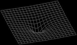
The cardiac muscle cells are organised in such a way that the conduction of the electrical waves through the heart go faster in one direction, called the fiber direction, than the other ones. This is comparable to the gps system that tells you, it will take 30 minutes to go from Kortrijk to Ghent when you take the highway instead of 50 minutes only using small roads. So we can redefine distance in terms of travelling time, instead of using the Euclidean distance.
In mathematics or physics terms, this comes down to endowing cardiac tissue with a metric tensor, and from geometric considerations, the heart then becomes a Riemannian manifold.
Using tensor calculus, geodesics and covariant derivatives, it is thereby possible to obtain general theoretical results on the time-evolution (drift and stability) of wave fronts and rotors in the heart. These efforts are laying the foundation for the field of “cardiac geometrodynamics”.
Application of mathematical physics concepts to cardiac excitation

Here is a non-exhaustive list of concepts from mathematical physics that are being used in our research:
- Geodesics, metric tensors, curved space
Anisotropy of wave propagation can be handled elegantly using a curved-space formalism. A glimpse thereof was added to a famous cardiology textbook. \ - Symmetry breaking
Wave fronts and rotors have less Euclidean symmetries than the reaction-diffusion equation, leading to critical eigenmodes of the linearized operator (Goldstone modes). - Bra-ket notation
In perturbation theory, we typically project onto the response functions, which can be written in Dirac’s notation: e.g. $\left<Y|PV\right>$. The use of quantum mechanical notation in biological context is sometimes confusing referees. - Particle-wave duality
In contrast to quantum mechanics, our operators are non-selfadjoint. As a result, the right-hand eigenfunctions are waves (spirals) while left-hand eigenfunctions are localized, like particles. This localization explains why it is so difficult to restore chaotic activity in the heart. See this great video. - Curved-space coordinate systems
In general relativity theory, it is customary to use nearly Euclidean coordinate systems, e.g. Gauss coordinates, Fermi coordinates or Riemann normal coordinates. We apply all of these in a biological context (and sometimes need to further extend them still). - Action principle
Part of the emerging rotor dynamics can be derived from an action principle. - Topological charge & defects
Cardiac rotors revolve around a rotor filament, which is a topological defect. We recently showed that the defect in 3D should be a phase defect surface. - String-like and brane-like dynamics
We previously showed that rotor filaments act as strings in a background space that is curved due to anisotropy. In the recent phase defect interpretation, filaments become brane-like objects that are phase defect surfaces. More topological constraints apply to the edges of those phase defect surfaces. - Green’s functions
Certain aspects can be dealt with classical superposition, e.g. forward calculation of electrograms and quantifying mechano-electrical feedback on rotor drift. - Branch cuts, complex analysis
Recent work shows that at the heart of a linear-core rotor, there is a phase discontinuity or phase defect. - Feynman-Hellman theorem
We use this theorem to calculate filament rigidity, which explains why in thin tissue slabs, full-fledged 3D instability cannot occur. - Pauli matrices and commutators
Even in three dimensions, rotation of scroll waves occurs in a plane, and the set of Pauli matrices is a suitable basis shape to calculate the shape of circular scroll wave cores, as well as the isotropic invariants of higher-order corrections. - Higher-dimensional embedding
We extended Wellner’s minimal principle for rotor filaments to inhomogeneous media by adding a fourth spatial dimension which restores homogeneity. - Schrödinger’s equation
The link between the diffusion and Schrödinger’s equation goes back a long time and has inspired the path integral formalism for quantum mechanics. Here, we explained paradoxical onset of ectopic (additional) heart beats using the analogy - in the other direction.
Building theory from experimental observations
To make the connection between theory and practice, we need to test our ideas on observations of excitation patterns in real hearts. However, these data are scarse, since it is not yet possible to view inside individual patients’ hearts.
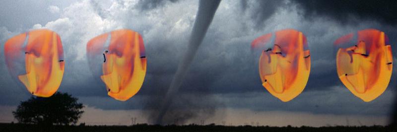
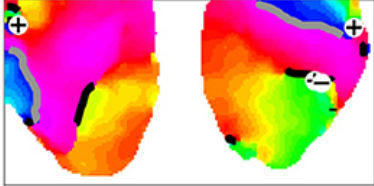
An experimental method that can be used on explanted hearts is optical voltage mapping. Here, a voltage-sensitive dye is administered to cardiac tissue to visualize excitation patterns with high resolution. Our first analysis within the group of arrhythmia patterns provided by Prof. E. Tolkacheva (Minneapolis, USA) demonstrated that cardiac rotors in rabbit hearts are organised around extended phase defect lines, rather than point singularities, forcing us to rethink the classical topological approach to cardiac arrhythmia organisation. In the next paper, we also identified the phase defects in cell cultures of human immortalized atrial myocytes, grown in the Pijnappels lab (University of Leiden, the Netherlands).
In ongoing work, we are performing pattern analysis and reconstruction on intracardiac electrograms, as well as ultrasound recordings.