Ultrasound imaging of arrhythmias
Even though various heart rhythm disorders can be recognized by electrocardiograms, the exact three-dimensional spatio-temporal pattern of the cardiac activation sequence throughout the cardiac wall is not well understood during rhythm disorders. Imaging these complex wave patterns can be done directly by plunging needle electrodes or needle catheters into the heart, however in human patients this is impossible to do and indirect inverse electrocardiographic techniques are being developed in order to infer the whole image from surface measurements. The best known example is the electrocardiogram (ECG), but to find the precise activation pattern from body-surface measurements is an incompletely solved inverse problem.
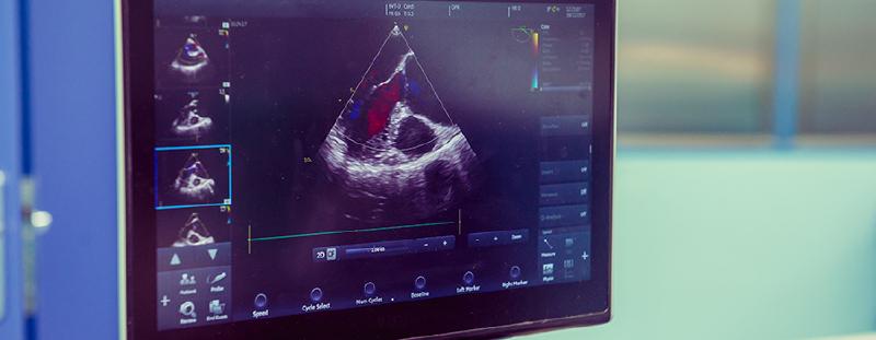
A pilot study from 2018 [1] showed that mechanical deformation of the heart, which can be estimated by ultrasound data, is closely linked to the electrical phenomena during cardiac arrhythmias. This idea opens up new ways of gaining insight into the complex, inherently 3D, electrical patterns in a fast and non-invasive manner. Recent studies have shown that imaging the electromechanical activation sequence with ultrasound data can be helpfull in certain situations (see for example in silico [2], in vivo experiments [3]).
In the ICARUS project, we set out to expand upon this technique and bring it to a clinically feasible tool, working in close collaboration the University Hospital UZ Leuven (Gasthuisberg). This collaboration involves the Cardiovascular Imaging and Dynamics group of Prof. Jan Dhooge and the group of Prof. Joris Ector, head of ablation therapies at the hospital. By combining expertise in mathematical modelling, echocardiography and clinical experience, we will advance our understanding of the three-dimensional electrical patterns and improve diagnosis and localization of cardiac arrhythmias.
[1] Christoph, J., Chebbok, M., Richter, C., Schröder-Schetelig, J., Bittihn, P., Stein, S., … Luther, S. (2018). Electromechanical vortex filaments during cardiac fibrillation, Nature, 555(7698), 667–672. https://doi.org/10.1038/nature26001
[2] Lebert, J., & Christoph, J. (2019). Synchronization-based reconstruction of electromechanical wave dynamics in elastic excitable media. Chaos, 29(9), https://doi.org/10.1063/1.5101041
[3] Grubb, C. S., Melki, L., Wang, D. Y., Peacock, J., Dizon, J., Iyer, V., … Wan, E. Y. (2020). Noninvasive localization of cardiac arrhythmias using electromechanical wave imaging. Science Translational Medicine, 12(536). https://doi.org/10.1126/scitranslmed.aax6111
Inversion of cardiac electrograms
When cardiac myocytes activate or deactivate, they act as electrical dipole sources, generating a potential field in the torso. This extracellular potential is recorded on the cardiac surface during surgery, or on the body surface, where it is known as the electrocardiogram (ECG). While the ECG is routinely used to diagnose and classify arrhythmias, it is still not possible to accurately reconstruct the arrhythmia sources in the heart from body-surface recordings.
As a step-up to ECG reconstruction, we aim to first solve the inverse problem for intracardiac electrograms (iEGM). From measurements with electrodes on the inner cardiac surface (endocardium), we seek to infer the 4D wave pattern inside the myocardial wall using physics-based inversion methods.
Cover image source: Griffin Health